
The Rise of 3D Technology in Neurosurgery
Introduction
Current technology has revolutionized different disciplines in the medical field such as neurosurgery. The development of 3D printing, 3D imaging, and HD imaging has brought the highest quality in imaging modalities to the field of neurosurgery. Minimally invasive brain surgery has become the standard of care in neurosurgery due to the use of endoscopic keyhole approaches coupled with high-quality 3D intra-operative imaging systems and 3D printing devices, thus improving the safety and quality of neurosurgery.
“3D printing has numerous potential applications in neurosurgery,” says Garni Barkhoudarian, MD, Director of the Skull Base and Endoscopic Microdissection Laboratory at Pacific Neuroscience Institute. “Customized, patient-specific surgical implants are currently available and are transforming the landscape in cranioplasty and the man-machine interface. Equally important is surgical education and disease modeling. A 3D printed specimen can train current and future surgeons to manage complex skull base diseases including invasive benign and malignant brain tumors”.
Medical facilities continue to use 2D imaging (CT and MRI) for treatment and surgical planning. Over the past several years, PNI has utilized 3D imaging for not only hands-on training purposes, but for patient education as well.
2D vs. 3D Imaging in Neurosurgery
Computer-Assisted Surgery (CAS) technologies such as neuro-navigation systems have enabled surgeons to perform intraoperative assessments of skull base and intra-axial tumors. These imaging systems are typically viewed in 2D (sagittal, axial, and coronal views). While 2D imaging is useful in term of navigation, 3D imaging provides the surgeon with a 360-degree, rotatable view. In terms of clarity, distortions of the anatomy may be evident in 2D while anatomical accuracy can be achieved in 3D. 2D systems produce a flat image while 3D imaging, is made up of multiple thin slices built into a volumetric 3D render. Moreover, 2D visual aids can take the form of photographs, illustrations, and clinical scans whereas 3D visual aids can be in the form of 3D medical imaging, computerized models, and volume-generated models from clinical scanning of 2D data.
Benefits of 3D Imaging
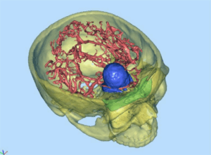
One benefit of 3D imaging is the ability to rotate the volume rendered model in real time for purposes of anatomy and diagnosis. 3D virtual reality models are volume-rendered from 2D CT (dicom – Digital Imaging and Communications in Medicine) images. Creating 3D interactive images in this way can improve visualization of structures and direct measurements can be made on 3D data sets, which can be limited on 2D images alone, thus enabling better medical decisions when viewing structures in the body. Additionally, 3D imaging enables easier tumor and anatomical location and eradicates the need for the viewer to interpret the depth of the image.
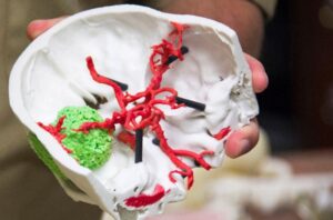
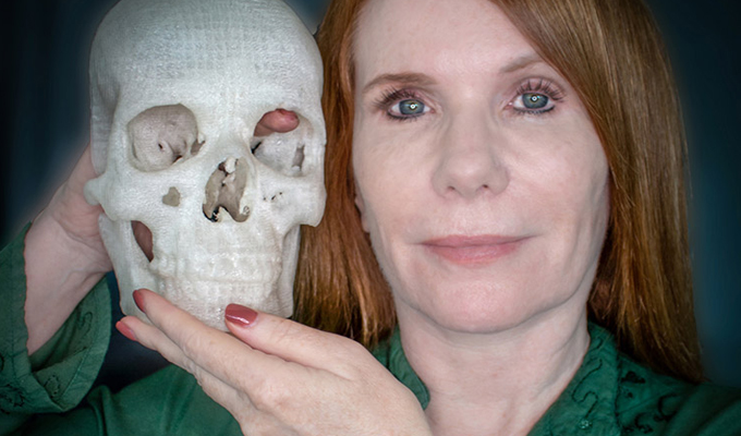
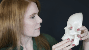
3D printed models enable the viewer to understand patient-specific anatomy in a comprehensive way. Once these images are rendered, 3D printers can produce models of the different anatomical structures in the human body thus enabling surgeons to have a clearer view of the anatomical structure. 2D imaging, on the other hand, produces “flat” images of anatomical structures requiring more skill from the surgeon to provide an accurate interpretation of the anatomy derived from the 2D images slices that include axial, coronal, and sagittal planes. Radiologists and surgeons may spot pathologies and anatomical discrepancies in 3D models that may potentially have been overlooked, thus advancing diagnosis and treatment planning.
3D Imaging in Advanced Medicine
In the early days of 3D printing for medical use in 2014, there was a great story about a man who saved his wife’s eyesight by 3D printing her brain tumor, a meningioma, which was getting entangled in her optic nerves. The 3D model convinced Pamela Shavaun Scott’s physicians to use a minimally invasive eyebrow keyhole approach to remove the tumor and save her vision.
Now many physicians around the world are practicing neurosurgical medicine utilizing a 3D approach to help diagnose and plan treatment. In fact, in March 2018, the 3D printing company Materialise became the first company to get FDA clearance for its 3D printing software to generate anatomical models for diagnostic use.
Some examples of the power of 3D include surgeons at Boston Children’s hospital who used a 3D printed brain to pioneer a medical breakthrough saving a toddler who suffered from terrible seizures known as “mind erasers”, giving him a normal life. And at the University of São Paolo’s Medical School in Brazil, neurosurgeons used a 3D printed brain model of an infant suffering from a rare congenital disorder called Sturge-Weber syndrome. The model was used both before and during the surgery as a guide for the surgeons during the successful operation to disconnect the part of the brain in which the infant’s seizures originated and save the infant’s life.
Patient Education
Explaining the anatomical structure of the brain, especially the cranial nerves, to patients using 2D diagrams can be challenging since patients may not fully grasp these concepts in a planar format. Physicians can now use 3D models to help describe intricate and complex anatomical structures such as a brain tumor, pituitary disorder, or cranial nerve compression, and how a surgery will be performed. Moreover, 3D models can be produced specific to the patient’s data set (CT or MRI), not only in 3D digital format, but as a 3D printed model as well.
For example, in the case of facial pain syndromes, during the patient visit, the physician can use a 3D model to more clearly explain the role of the cranial nerves and specifically discuss the particular issues experienced by the patient. In many cases, a microvascular decompression procedure to help alleviate symptoms could be discussed with the help of the 3D model based on the patient’s own imaging.
Neurosurgical Skills Skull Base and Endoscopic Microdissection Laboratory
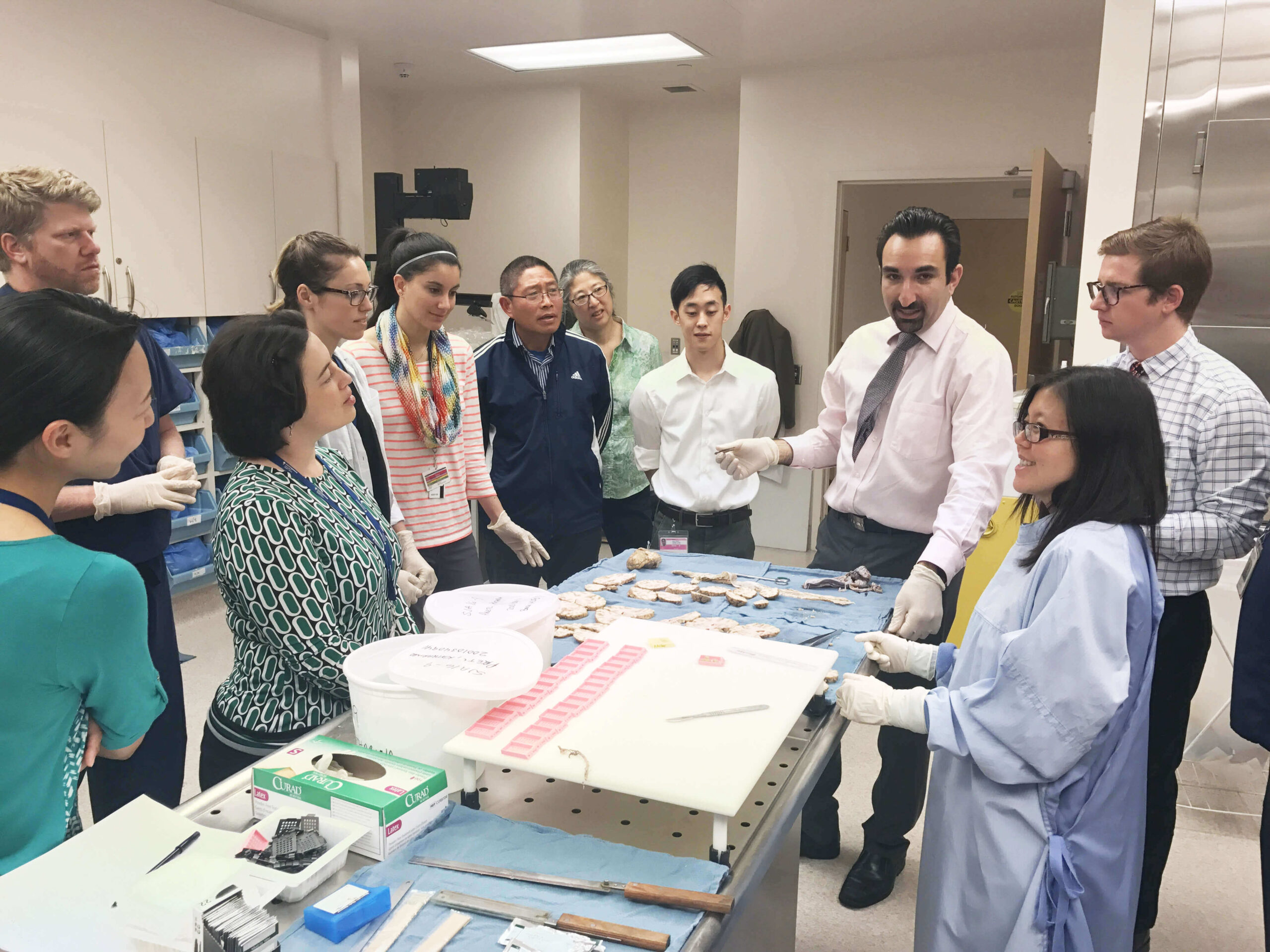
At Pacific Neuroscience Institute, the neurosurgical skills skull base and microdissection lab is utilized by fellows, visiting scholars, medical students, and faculty alike to practice neurosurgical procedures. In addition to the use of cadavers, 3D printing is used to recreate multiple neurosurgical pathologies for a more accurate experience in the neurosurgical field. The multidisciplinary team can educate colleagues and help them practice endoscopic keyhole approaches on 3D printed anatomical models.
Conclusion
3D imaging is changing the future of medicine. With more developments in technology in areas such as artificial intelligence and computational power, image acquisition and processing times will be greatly reduced. Additionally, medical imaging data will likely be cloud-hosted freeing up local computer storage which is considerable for 3D renders. Along with 2D imagery, 3D models provide a more complete picture of the patient’s condition and are another tool that neurosurgeons can use to diagnose and plan treatment for difficult neurological diseases.
For more information about minimally invasive brain surgery and advanced surgical techniques, contact us at the Pacific Brain Tumor Center at 310-582-7450.

Mahyar Pakizegee is a fourth-year baccalaureate student at the University of California, Los Angeles majoring in History. His goal is to attend medical school and pursue his dream of being an inspiring physician. He is a contributing author under the guidance of Dr. Garni Barkhoudarian, neurosurgeon at Pacific Neuroscience Institute.

Nicolette Mena is the PNI Foundation Program Coordinator and is involved with all administrative and operational aspects of the Pacific Neuroscience Institute Foundation. She focuses on raising awareness of PNI, through composition of blog posts, video appeals, newsletters, and materials for the bi-annual magazine. Fundraising is Nikki’s priority, with her efforts geared toward grant writing and coordination of outreach events. She works closely with medical experts, Saint John’s Health Center Foundation’s development team and PNI Foundation’s Directors to expand PNI’s brand both domestically and internationally.