Focused Ultrasound
What is Focused Ultrasound?
Focused Ultrasound is an incisionless treatment for patients with Essential Tremor (ET) and Tremor Dominant Parkinson’s Disease (TDPD) who have not responded to medication. It uses high intensity ultrasound waves guided by MRI to treat deep areas in the brain associated with tremor, with no permanent implants or general anesthesia. There is little to no risk of infection and patients usually return home the same day with immediate tremor improvements.
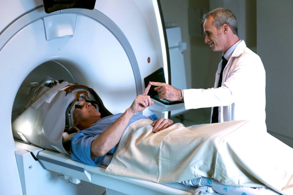
Focused ultrasound targets a small brain area called the ventral intermediate nucleus, which is a crucial relay station for tremor-related signals. This precise targeting is a crucial aspect of its efficacy in reducing tremors. During the procedure, the patient is lying on a table and moves in and out through an MRI. The MRI enables the physician to visualize a patient’s brain anatomy, plan, and target the area for treatment. It also acts like a thermometer, providing continuous temperature monitoring to verify that only the targeted tissue is destroyed. As the temperature at the brain target rises, it creates a small ablation or burn, providing a therapeutic effect.
While treatment of hand tremors is the goal of focused ultrasound, it may also provide some degree of tremor relief for the head and voice.
Medical Indications
- Medication-Refractory Tremor: Patients with ET or TDPD who have not adequately responded to medication may be considered.
- Severe Tremor Impacting Quality of Life: Candidates typically have tremors that significantly affect their daily activities, work, and social interactions, thereby impairing their quality of life
- Patient Preference: Particularly suitable for patients who prefer less invasive procedures and patients who want to avoid the upkeep of an implantable device.
- Contraindications for Surgery: Patients who cannot undergo surgical procedures due to medical contraindications may be eligible, as it avoids surgical risks such as infection and bleeding.
- Treatment of Unilateral Symptoms: The treatment is FDA approved for staged unilateral treatment – treating one side at a time with at least 9 months between treatments for eligible patients and without significant clinical events from the first treatment.
- Assessment and Diagnosis: Patients are typically assessed to ensure that their tremor symptoms are primarily caused by ET or TDPD and no other neurological conditions, ensuring accurate diagnosis and treatment.
Contact us to learn more: Call 310-961-4024 or Request an Appointment.
Focused Ultrasound Specialists


Written and reviewed by:
The Pacific Neuroscience medical and editorial team
We are a highly specialized team of medical professionals with extensive neurological and cranial disorder knowledge, expertise and writing experience.
Last Updated: