Hemangioblastoma
Team
Meet our Expert Specialists & Surgeons


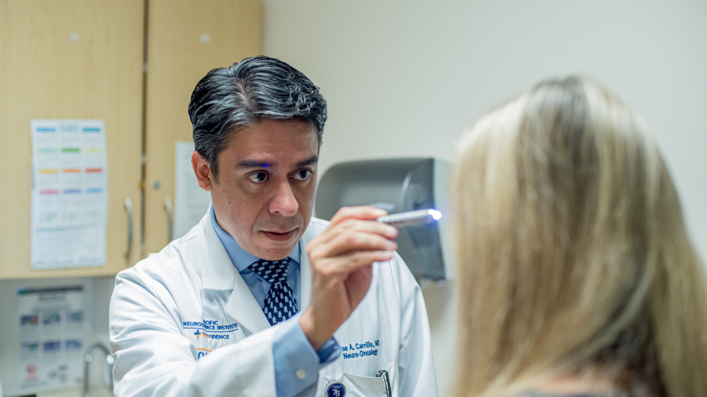
Care at PNI
Experience Compassionate, Expert Care

Written and reviewed by:
The Pacific Neuroscience medical and editorial team
We are a highly specialized team of medical professionals with extensive neurological and cranial disorder knowledge, expertise and writing experience.
Last Updated: