Craniopharyngioma: The Most Formidable of Intracranial Tumors
Originally published November 14, 2017 | Updated August 27, 2026
Treatment Challenges and Advances
In 1939, the father of modern neurosurgery, Dr. Harvey Cushing, called craniopharyngiomas “the most formidable of intracranial tumors.” At the time, surgeons had no microscope, no endoscope, and none of today’s hormone replacement therapies to help patients recover from surgery near the pituitary gland.
Cushing’s description still holds true.
Craniopharyngiomas remain some of the most complex tumors to treat safely. What has changed is our ability to treat them: modern endoscopic surgery, targeted radiation, and hormone replacement have transformed outcomes for patients of all ages.
This article walks through how our team diagnoses and treats craniopharyngiomas today, from initial symptoms through long-term follow-up. For a complete overview of causes, symptoms, and treatment options, see our craniopharyngioma condition guide.
Overview
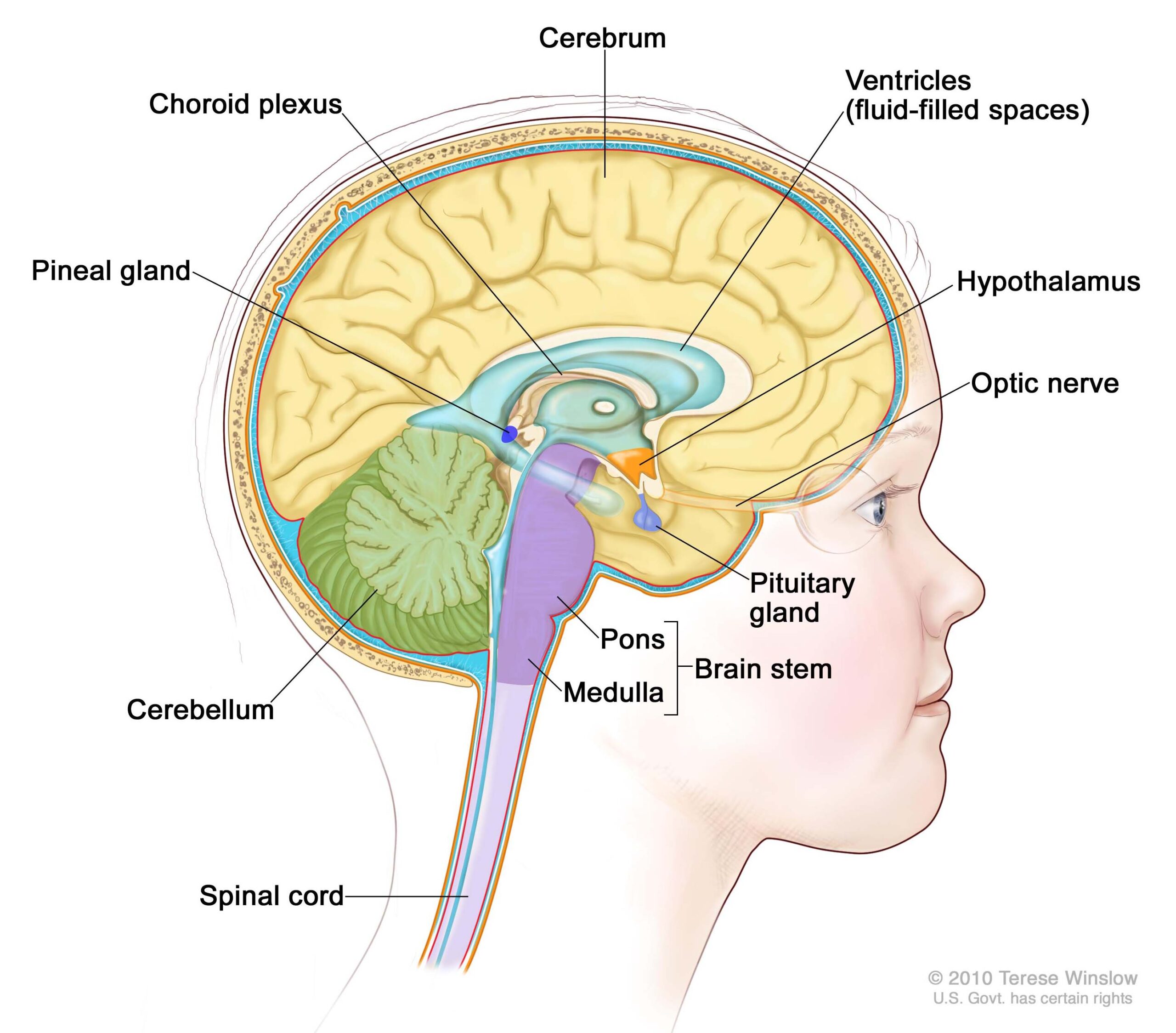
Although classified as benign (non-cancerous), craniopharyngiomas pose a great challenge in management because they typically adhere to the pituitary gland and stalk, optic nerves and optic chiasm, surrounding blood vessels as well as the brain itself.
- They are thought to arise from developmental remnants of the craniopharyngeal duct or Rathke’s pouch.
- They can occur at any age but are most common in childhood and adolescence and in adults over age 50.
- They account for 10-15% of sellar and suprasellar tumors (tumors that occur in and above the pituitary gland) and over 50% of such tumors in children.
- They are usually not discovered until they impinge upon important structures, and are frequently quite large (over 3 cm) when detected.
Symptoms
The most common symptoms of craniopharyngiomas are related to loss of hormonal function (hypopituitarism), including low energy, low libido, loss of menstrual periods in women and weight gain, growth failure in children), loss of vision, and headache.
Some craniopharyngiomas may also become quite large, blocking flow of cerebrospinal fluid (CSF), leading to hydrocephalus with changes in mental state and cognition.
Diagnosis
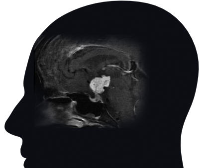
Craniopharyngiomas are typically diagnosed on MRI or CT scan, and are usually distinguishable from the more common pituitary adenomas, Rathke’s cleft cysts, and meningiomas that might occur in the region around the pituitary gland and skull base.
Craniopharyngiomas typically have both solid and cystic components, and many show calcification on CT scan.
Complete pituitary hormonal testing is essential given that a majority of patients have hormonal deficits. Most patients will also need formal visual field and acuity testing to assess the tumor’s impact on vision and eye movements.
Treatment
First-line treatment for craniopharyngiomas is maximal safe surgical removal, although in a minority of mostly cystic craniopharyngiomas, simple cyst drainage may be the first treatment. Second-line treatment is typically focused radiotherapy or radiosurgery.
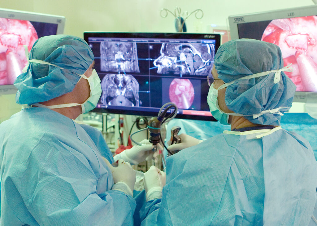
Given that most craniopharyngiomas arise along the pituitary stalk, the most common surgical approach is the endoscopic endonasal route via the nostrils. If pituitary gland function is intact, every attempt is made to preserve the integrity of the pituitary gland, stalk, and its connection to the hypothalamus. Alternatively, for tumors that extend far off the midline and for some recurrent craniopharyngiomas, a supraorbital eyebrow craniotomy can be used1.
Given the tendency of craniopharyngiomas to adhere to critical surrounding blood vessels, cranial nerves and the brain itself, particularly the hypothalamic region, “near complete” or subtotal removal occurs in a majority of patients while complete (100%) removal occurs in less than half of cases. This approach also has a higher likelihood of preserving pituitary gland function. Typically, if small bits of tumor are left behind, these are closely monitored, and then treated with focused stereotactic radiotherapy or radiosurgery, if they grow on subsequent MRI.
This treatment formula of maximal safe surgery followed by focused radiation if the residual tumor grows, has a high success rate in our hands as well as at other high-volume Pituitary Centers of Excellence. A relatively new discovery is that a subset of craniopharyngiomas described as “papillary” typically have a BRAF mutation that can be a target for a BRAF inhibitor drug, now the focus of a clinical trial for patients whose tumors are not controlled with surgery and radiation.
Follow-Up
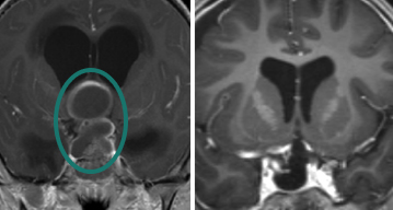
Craniopharyngioma patients need long-term neurosurgical and endocrine follow-up with regular MRIs and hormonal evaluations, as late recurrences can occur.
We typically follow our patients with regular MRIs every 3-6 months for at least 2-3 years post-surgery and then annually for at least 10 years.
PNI’s Experience with Craniopharyngiomas
With an overall series of over 2,000 endonasal operations for all types of pituitary and related skull base tumors, our Pacific Pituitary Disorders Center team has a long track record treating these highly complex tumors using a team approach of neurosurgery, ENT, endocrinology, radiation oncology, neuro-ophthalmology, and neuro-oncology.
Since 2009, we have used an exclusively endoscopic endonasal approach for all pituitary tumors including craniopharyngiomas, given the optimal visualization the high-definition endoscope affords of the pituitary gland and related skull base anatomy.
In our experience of craniopharyngioma surgeries using our fully endoscopic technique, 80 percent2 of patients undergoing first-time surgery achieved a near-total or gross-total tumor removal with a very low complication rate and a high rate of gland and stalk preservation. Our typical endonasal craniopharyngioma surgery takes about 5 to 6 hours, and most patients are discharged home the day after surgery.
FAQs about Craniopharyngiomas
Is a craniopharyngioma a type of brain cancer?
No, they are not considered a cancer (malignant). Craniopharyngiomas are brain tumors that are classified as benign, meaning they do not spread to other parts of the body. Because they grow close to the pituitary gland, optic nerves, and brain itself, they can still cause serious health problems and require expert treatment.
What is the recommended treatment for craniopharyngioma?
The first line of treatment is maximal safe surgical removal, most often through endoscopic endonasal surgery via the nostrils or, in select cases, a supraorbital eyebrow craniotomy. If any tumor remains after surgery, focused radiation is typically used to control any residual growth.
Can a craniopharyngioma come back after surgery?
Yes. Recurrence is possible even years after treatment, which is why patients need long-term monitoring with MRI and hormone testing, typically every 3 to 6 months for the first several years and then annually for at least a decade.
Do patients with craniopharyngioma need hormone replacement for life?
Many do. Because craniopharyngiomas grow near the pituitary gland, most patients develop some degree of hormone deficiency from the tumor itself, surgery, or radiation, and require ongoing hormone replacement monitored by an endocrinologist.
Who treats craniopharyngioma at Pacific Neuroscience Institute?
Craniopharyngioma care requires a team approach. At PNI, that team includes neurosurgeons, ENT skull base surgeons, endocrinologists, radiation oncologists, and neuro-ophthalmologists working together on diagnosis, surgery, and long-term follow-up.
What is the outlook for someone diagnosed with a craniopharyngioma?
With expert surgical and hormonal care, most patients achieve good long-term tumor control and quality of life, though lifelong follow-up is generally required to watch for recurrence and manage hormone levels.
Citations
- The Supraorbital Eyebrow Craniotomy for Intra- and Extra-Axial Brain Tumors: A Single-Center Series and Technique Modification. https://pubmed.ncbi.nlm.nih.gov/32745195/
- Endonasal versus supraorbital keyhole removal of craniopharyngiomas and tuberculum sellae meningiomas. https://pubmed.ncbi.nlm.nih.gov/19287324/
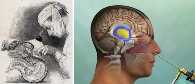
Banner image: Dr. Harvey Cushing performing sublabial transsphenoidal surgery with a head lamp, drawn in 1912 by Max Brodel, contrasted with current endonasal transsphenoidal approach for craniopharyngioma illuminated with high definition endoscope. Craniopharyngioma endoscopic endonasal approach ©Pacific Neuroscience Institute.
About the Author