Acoustic Neuroma
At Pacific Neuroscience Institute, our experts are focused on compassionate, state-of-the art, multidisciplinary care for patients with acoustic neuroma and other schwannomas.
On this page:
Overview | Symptoms | Diagnosis | Treatment | FAQs
Expanded Care | Doctors | Videos
Clinic Locations | Patient Resources
Overview of Acoustic Neuroma
Acoustic Neuroma (AN, vestibular schwannoma) is a relatively rare, benign (noncancerous) skull base tumor affecting 1 in 100,000 people. The only definitive environmental risk factor that has been associated with acoustic neuroma is radiation exposure of the whole head. Acoustic neuromas can be related to a genetic syndrome called neurofibromatosis, and bilateral acoustic neuromas (affecting both ears) are associated with neurofibromatosis type 2 (NF2).
Acoustic neuromas arise from an over-production of Schwann cells of the nerve (sheath) covering of the eighth cranial nerve (the vestibulo-cochlear nerve). This nerve runs from the inner ear to the brain and governs hearing and balance.
While each patient tends to experience acoustic neuroma differently, the most common symptom is hearing loss in one ear followed by tinnitus (ringing). As the tumor enlarges, it compresses the nerve often causing dizziness, imbalance, and coordination difficulties. In addition, other symptoms may include numbness, tingling, and difficulty swallowing. If the tumor is very large facial weakness or paralysis may occur.
Our otology (hearing) speciality surgeons and neurosurgeons personalize treatment for each patient. If surgery is indicated, minimally invasive keyhole and endoscopic surgery is available for most symptomatic acoustic neuromas.
Acoustic Neuroma Naming Conventions
Acoustic neuroma may be referred to by several terms.
- Acoustic neuroma
- Acoustic neurilemoma
- Acoustic neurinoma
- Fibroblastoma, perineural
- Neurinoma of the acoustic nerve
- Neurofibroma of the acoustic nerve
- Schwannoma of the acoustic nerve
- Vestibular schwannoma
Acoustic Neuroma Symptoms
Patients tend to experience symptoms in accordance with the size of the tumor, although that in not always the case. In general, smaller acoustic neuromas cause fewer symptoms than larger ones.
Acoustic neuromas grow on the vestibulo-cochlear nerve which is responsible for balance. The nerve leads from the inner ear to the brain in an area called the cerebellopontine angle. As the tumor grows, it may protrude into the internal auditory canal, which also contains the facial nerve, cochlear (auditory) nerve, and the vestibular (balance) nerves. Very large tumors compress the auditory canal, cerebellum, and brainstem, and are considered to be life threatening.
Based on tumor size and compression severity, acoustic neuroma symptoms include:
- Hearing loss in one ear
- Tinnitus (ringing)
- Dizziness
- Imbalance
- Coordination difficulties
- Facial numbness
- Facial tingling
- Difficulty swallowing
- Facial weakness or paralysis may occur with very large tumors
Acoustic Neuroma Diagnosis
Acoustic neuromas are typically diagnosed by an MRI with gadolinium or a CT scan of the brain. A focused MRI of the internal auditory canals is typically best for visualizing acoustic neuromas.
Other tests may also be needed such as:
- Angiography (typically now performed as a CT angiogram or an MR angiogram)
- Audiograms
Acoustic Neuroma Treatment
At Pacific Neuroscience Institute, our experts are focused on compassionate, state-of-the art, multi-specialty care for patients with acoustic neuromas. We are focused on your health and our team encourages shared discussion of treatment strategies. Your treatment team considers all aspects of disease presentation to provide a comprehensive, personalized, management plan.
Patient treatment falls into three categories:
- Observation
- Radiosurgery and fractionated radiation
- Minimally invasive surgery
Observation
Our conservative course of action is regular observation for patients who have small tumors (less than 2 cm) and who are experiencing few symptoms that are not affecting hearing or quality of life. This involves a second MRI 6 months after diagnosis, followed by annual imaging to monitor tumor size, growth, and symptoms. These tumors may appear to remain unchanged over many years but It is important to note however that hearing loss often progresses even though small tumors may not show any growth.
Radiosurgery and Fractionated Radiation
For larger or symptomatic tumors that require a more aggressive approach, patients may receive advanced-technology focused radiosurgery or stereotactic radiotherapy, or fractionated radiation which targets tumor tissue while sparing the surrounding areas of healthy brain. This nonsurgical treatment is typically done in an outpatient setting where radiation may be administered as a single dose or in multiple or fractionated doses.
Minimally Invasive Neurosurgery
For those who may be candidates for minimally invasive surgery, our team of experienced surgeons use keyhole and endoscopic approaches to make acoustic neuroma and schwannoma surgery safer, less invasive, and more effective to optimize your clinical outcomes.
For tumors under 2.5 cm, either surgery or radiosurgery are reasonable treatment options. For tumors over 2.5 cm, surgical removal is generally recommended.
Treatment for acoustic neuromas by surgical removal is through a keyhole retrosigmoid craniotomy, a middle cranial fossa, or a translabyrinthine minimally invasive route using an endoscope and micro-instrumentation. The choice of approach depends upon the tumor location. Tumor adherence to critical structures dictates how much tumor can be safely removed, and in some cases, not all tumor tissue can be removed. In some patients in whom only a sub-total tumor removal is possible through neurosurgery, radiosurgery or stereotactic radiotherapy may be effectively used to control further tumor growth.
- The retrosigmoid approach to treat acoustic neuroma (also known as the retromastoid approach) uses a small window behind the ear to reach and remove the acoustic neuroma. This approach is augmented with the use of the endoscope, resulting in minimal cosmetic or soft-tissue damage and relatively quick patient recovery. In this approach the inner ear structures can remain intact to preserve hearing. Patients with functional hearing and small tumors (under 2 cm) that do not extend deep into the internal auditory canal can benefit from this approach. For patients with a history of headaches we avoid retrosigmoid surgery as there is a low risk of chronic headache with this approach.
- The middle cranial fossa surgical approach for acoustic neuroma involves a small opening above the ear for small tumors (less than 1.7 cm) that are typically confined to the internal auditory canal. This procedure aims for long-term hearing preservation in patients with functional hearing. Using this minimally invasive approach, the neuroma can be reached from above without disturbing the structures of the inner ear.
- The translabyrinthine approach for acoustic neuroma is performed through a small incision behind the ear. This treatment modality offers the most direct route and widest view of the skull base. It is the preferred approach for larger tumors (greater than 2 cm) where hearing loss has progressed or where hearing preservation is unlikely.
The benefits of minimally invasive surgery compared to open surgery are:
- Fewer headaches
- Less disruption to surrounding brain tissue
- 1-2 nights in the hospital
- Less disruption to patient’s day to day life
Acoustic Neuroma FAQs
Is acoustic neuroma serious?
Some acoustic neuromas can grow very large and cause major brain / brainstem compression which could affect major neuroogical problems over time.
What are the symptoms of acoustic neuromas?
Acoustic neuromas can cause hearing loss, facial numbness, pain or weakness, headaches, imbalance, swallowing difficulties and weakness.
How is acoustic neuroma treated?
Large acoustic neuromas are treated with surgical resection. Small acoustic neuromas can be treated with surgery or stereotactic radiosurgery, though some can be observed to see if they may grow or cause neurological deficits.
Are acoustic neuromas painful?
Some can cause headaches and/or facial pain.
Can acoustic neuromas spread?
No, acoustic neuromas don’t spread.
How is acoustic neuroma diagnosed?
Acoustic neuromas are diagnosed by MRI and audiogram.
How do you treat a nerve sheath tumor?
A nerve sheath tumor is treated by surgical resection (removal).
Are acoustic neuromas benign?
Yes, most often acoustic neuromas are benign (noncancerous).
Is acoustic neuroma a brain tumor?
Yes, acoustic neuroma is considered a brain tumor.
Are acoustic neuromas cancerous?
Acoustic neuromas are not cancerous.
Can acoustic neuromas cause facial paralysis?
Yes, acoustic neuroma patients are at risk for facial nerve paralysis. Factors include size of tumor, location, and growth rate. Some patients may develop facial paralysis prior to diagnosis/treatment due to compression of the facial nerve. If the facial nerve is injured as a result of removal of the acoustic neuroma, patients may experience facial paralysis after surgery and it is essential to consult a facial reanimation expert immediately and create a plan to manage the paralysis.
Expanded Care for Acoustic Neuroma
You will be supported by our expanded team including experts in facial reanimation, neurology, hearing and balance, physical therapy and rehabilitation, and clinical neuropsychology.
Facial Nerve Paralysis Treatment for Acoustic Neuroma
In some patients with AN, facial paralysis may develop prior to diagnosis/treatment due to compression of the facial nerve. This can be because of the size and/or location of the tumor since the facial nerve (cranial nerve 7) and 8th cranial nerve are located very close together. If the acoustic neuroma has caused facial nerve damage resulting in facial paralysis, our facial reanimation and plastics reconstructive surgeon is also included in the treatment team. Treatment options for AN-related facial paralysis may vary and are dependent on the health of the facial nerve. We recommend a consultation with our facial reanimation expert right after surgery to create a plan to manage the paralysis.
Vestibular Rehabilitation (Balance Therapy)
Balance therapy is an integrated component of your care. Vestibular status is assessed before and after your procedure by physical therapists with expertise in issues related to balance and dizziness. Our vestibular rehabilitation program is composed of a series of balance training exercises that are individually tailored for each patient. These exercises promote the patient’s vestibular system to compensate for the balance disorder. Our specialists work in conjunction with the Performance Therapy department at Providence Saint John’s Health Center to provide balance therapy services to our patients.
Other Services
- Speech and swallowing
- Psychosocial support
- Audiology
Acoustic Neuroma Doctors and Specialists
Clinic Locations
Pacific Neuroscience Institute – Santa Monica
2125 Arizona Ave, Santa Monica, CA 90404
310-829-8701
Pacific Neuroscience Institute – Wilshire
11645 Wilshire Blvd, #600, Los Angeles, CA 90025
310-477-5558
Acoustic Neuroma Specialists & Videos
 Meet Dr. Courtney Voelker
COURTNEY C.J. VOELKER, MD, PhD Director, Otology/Neurotology-Lateral Skull Base Surgery; Director, Adult & Pediatric Cochlear Implant Program Dr. Voelker is an otolaryngology – head & neck surgeon who takes care…
 A PNI Minute | Acoustic Neuroma with Dr. Courtney Voelker
Dr. Courtney Voelker explains about acoustic neuroma, a benign tumor that affects the nerve for hearing and balance. Find out about signs and symptoms, and the three treatment options that…
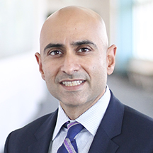 Meet Dr. Amit Kochhar
Dr. Amit Kochhar, MD, is double board-certified in Otolaryngology, Head and Neck Surgery and Facial Plastic and Reconstructive Surgery. He is the director of the Facial Nerve Disorders Program at…
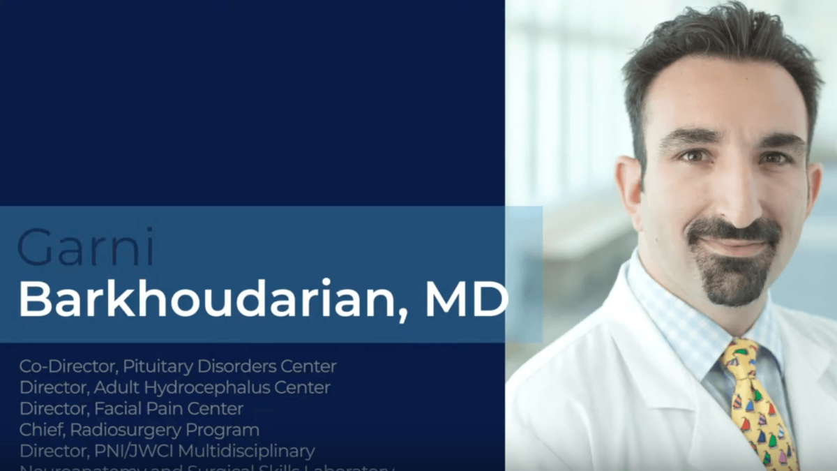 Meet Dr. Garni Barkhoudarian
Dr. Barkhoudarian is the Co-Director Pituitary Disorders Center, Neurosurgery; Director, Adult Hydrocephalus Center; Director, Facial Pain Center; Chief, Radiosurgery Program; Director, JWCI / PNI Microdissection Anatomy Laboratory. He is a…
 Meet Dr. Daniel F. Kelly
 Meet Dr. Walavan Sivakumar
Dr. Walavan Sivakumar is a fellowship-trained neurosurgeon with a focus on skull base and minimally invasive and endoscopic neursosurgery. Selected as a multiple year recipient of the SuperDoctor Rising Stars…
 Meet Dr. Adi Iyer
 Meet Dr. Nouzhan Sehati
Dr. Sehati, is a board-certified neurosurgeon who specializes the treatment of a wide variety of brain, spine and spinal cord disorders at Pacific Neuroscience Institute. His expertise is in surgical…
 Meet Dr. Gowrinathan
Dr. Gowrinathan is the director of Psycho-oncology, Brain Health Center; Psychiatry, Psycho-oncology. She is an accomplished inpatient and outpatient psychiatrist, specializing in both Women’s Psychiatry and Psycho-oncology (Cancer Psychiatry) who…
 Meet Dr. Rebecca Lewis
REBECCA LEWIS, AuD Audiology Director, Adult & Pediatric Cochlear Implant Program; Audiologist Dr. Lewis is a board certified audiologist focusing on adult and pediatric patients. An avid clinician, educator, and…

Meet Dr. Courtney Voelker

A PNI Minute | Acoustic Neuroma with Dr. Courtney Voelker

Meet Dr. Amit Kochhar
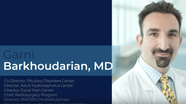
Meet Dr. Barkhoudarian

Meet Dr. Daniel F. Kelly

Meet Dr. Walavan Sivakumar

Meet Dr. Adi Iyer

Meet Dr. Nouzhan Sehati

Meet Dr. Gowrinathan


