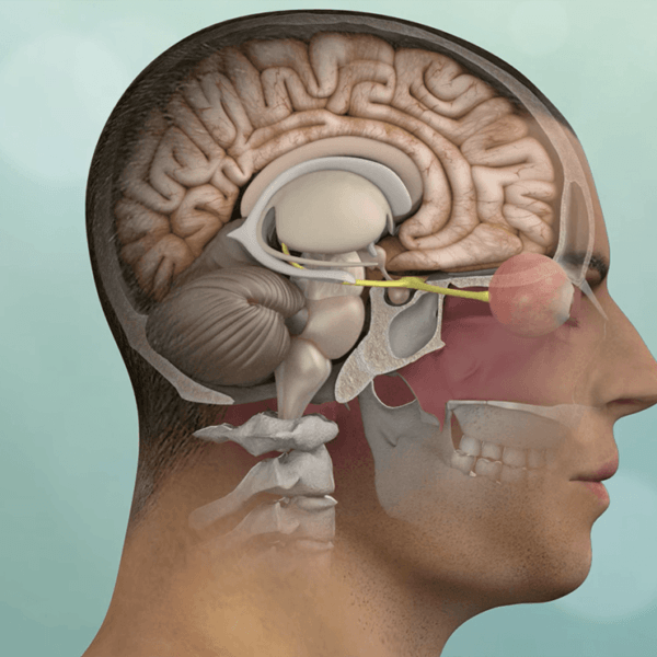
Colloid Cyst
What is a Colloid Cyst?
Colloid Cysts are benign cystic fluid collections that occur within the fluid-filled ventricles of the brain.
Colloid cysts develop in the brain at the junction of the paired lateral ventricles and can cause blockage of cerebrospinal fluid (CSF) flow leading to hydrocephalus (excess brain CSF). As a result, these benign growths can cause headaches, visual changes, memory difficulties and occasionally result in loss of consciousness or coma.
Fortunately, most symptomatic or large colloid cysts can now be safely removed through a minimally invasive endoscopic technique or brain port technique via a quarter-sized bony opening in the skull. This procedure typically resolves the hydrocephalus and associated symptoms.
These relatively uncommon benign cysts arise within the fluid filled regions of the brain, the ventricles. They typically occur at the junction between the lateral and third ventricles. The cyst consists of a thin lining surrounding a thick fluid-filled center. Once they reach a critical size, these cysts can block the normal flow of cerebrospinal fluid (CSF), increasing the pressure within the brain. Additionally, they can compress the nervous structures that process memory signals in the brain.
Symptoms of Colloid Cyst
As slow growing colloid cysts enlarge, they start to obstruct the normal flow of CSF through the Foramina of Munro which are the narrow channels through which CSF flows from the two lateral ventricles into the 3rd ventricle. This obstruction can result in increased pressure in the head leading to:
- Headaches
- Vision disturbance
- Double vision
- Memory and concentration difficulties
- In some advanced cases, altered states of consciousness and even coma.
Diagnosis of Colloid Cysts
Colloid cysts are typically diagnosed by magnetic resonance imaging (MRI) or computer tomography (CT) scans of the brain. Specific high resolution MRI sequences can further characterize colloid cysts, how they impact surrounding normal anatomical structures and CSF flow patterns from the lateral into the 3rd ventricle.
Colloid Cyst Surgery Treatment
For symptomatic colloid cysts, the best treatment is surgical removal. Depending on the exact anatomical location of the colloid cyst and the size of the lateral ventricles, surgical options include endoscopic resection of the colloid cyst or use of a brain port for a minimally invasive transcranial resection of the colloid cyst. In rare cases, ventricular drains (aka VP shunts) may be necessary after surgery to divert CSF around the blocked Foramina of Munro.


