Shunts
Types of Shunts
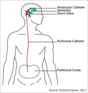
This common procedure is performed with a “GPS” neuronavigation system to ensure accurate catheter placement. There are a variety of valves available, though we prefer one of a few programmable valves. These valves may have an MRI lock system to prevent unwanted shunt adjustments in a magnetic field. Some have “Off” switches to determine the necessity of the shunt without requiring to perform a surgical procedure to tie it off.
The abdominal portion of the catheter is placed laparoscopically with the assistance of a general surgeon, ensuring correct placement of the catheter and minimizing the risk of abdominal organ injury.
Though a common procedure, this device can have some complications during and after placement. These include catheter misplacement, catheter dislodgement, valve obstruction, shunt infection, abdominal organ injury, overdrainage, possible revision surgery or removal surgery and intracranial bleeding (acute or delayed).
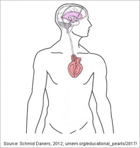
This procedure is also well tolerated and carries similar risks to a VP shunt. Though the risk to the abdominal organs is eliminated, there is a slightly higher risk of bloodstream infections or heart valve infection (endocarditis). Also, there is a higher risk in patients with cardiac arrhythmias as the catheter can irritate the heart rhythm control region.
Placing a new VA shunt requires an inpatient stay. However, converting from a VP to VA shunt can be done on an outpatient basis for some patients.
This device can have some complications during and after placement. These include catheter misplacement, catheter dislodgement, valve obstruction, shunt infection, overdrainage, possible revision surgery or removal surgery and intracranial bleeding (acute or delayed). These shunts are less durable than VP shunts and can require more frequent revision operations.
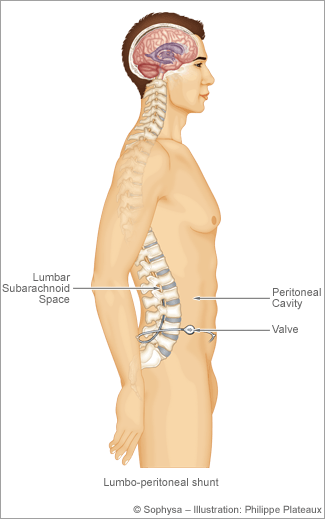
The abdominal portion of the catheter is placed laparoscopically with the assistance of a general surgeon, ensuring correct placement of the catheter and minimizing the risk of abdominal organ injury.
Though a common procedure, this device can have some complications during and after placement. These include catheter misplacement, catheter dislodgement, valve obstruction, shunt infection, overdrainage, possible revision surgery or removal surgery and intracranial bleeding (acute or delayed).

Written and reviewed by:
The Pacific Neuroscience medical and editorial team
We are a highly specialized team of medical professionals with extensive neurological and cranial disorder knowledge, expertise and writing experience.
Last Updated: