Benign Sinonasal Tumors
Diagnosis
Benign Sinonasal Tumor Diagnosis
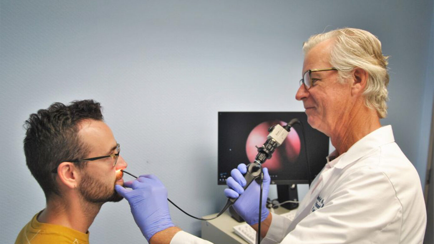
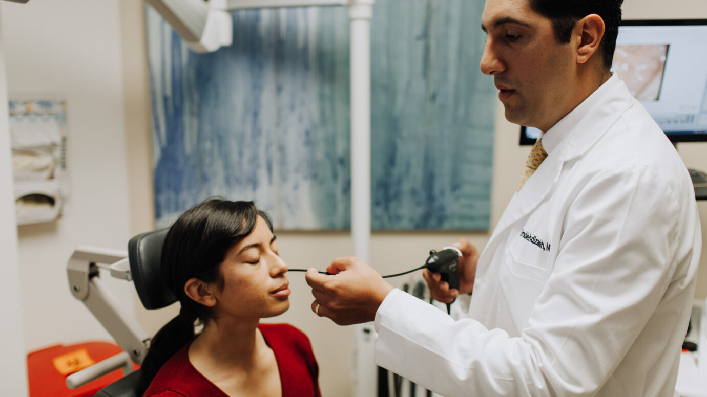
Diagnosis of benign sinonasal tumors usually involves a combination of:
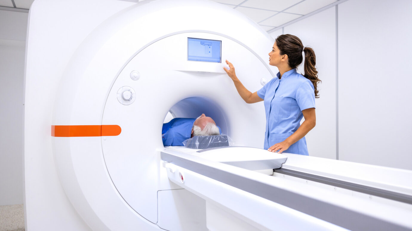

In some patients these tumors are found incidentally on imaging for other reasons.


Care at PNI
Experience Compassionate, Expert Care

Written and reviewed by:
The Pacific Neuroscience medical and editorial team
We are a highly specialized team of medical professionals with extensive neurological and cranial disorder knowledge, expertise and writing experience.
Last Updated: