Arteriovenous Malformation
What is Arteriovenous Malformation?
Overview
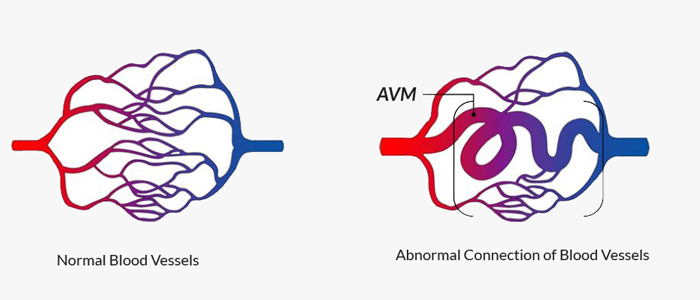
An arteriovenous malformation (AVM) is a vascular mass made up of tangled blood vessels in which one or more direct connections form between arteries and veins.
Normally, arteries are strong, thick-walled tubes that carry high-pressure blood from the heart to the brain and body. Veins, on the other hand, are thinner and carry low-pressure blood back to the heart. In an AVM, blood flows directly from arteries to veins, bypassing the normal capillary system. This exposes delicate veins to high-pressure arterial blood flow, making them prone to rupture, which can lead to bleeding in the brain (hemorrhage).
These abnormal blood flow patterns may also cause weakening of nearby arteries and increase the risk of aneurysm formation.
Who is Affected?
AVMs are relatively rare, affecting less than 1% of the population. They can occur anywhere in the brain or spinal cord and are often present from birth, although symptoms may not appear until later in life. Most AVMs are diagnosed in young adults between ages 20 and 40, but they can also be found in children or older adults. Men and women are affected at similar rates.
Arteriovenous Malformation Diagnosis
Diagnosis typically begins with brain imaging. These studies help determine the size, location, and flow characteristics of the AVM, which guide treatment planning. They include:


Meet our Expert Specialists & Surgeons


Experience Compassionate, Expert Care


Written and reviewed by:
The Pacific Neuroscience medical and editorial team
We are a highly specialized team of medical professionals with extensive neurological and cranial disorder knowledge, expertise and writing experience.
Last Updated: