Brain Aneurysm
What is a Brain Aneurysm?
Overview
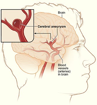
A brain aneurysm—also called a cerebral aneurysm or intracranial aneurysm—is a weakened, bulging area in the wall of an artery that supplies blood to the brain. Arteries are vital blood vessels that carry oxygen-rich blood from the heart to the brain, nourishing brain cells and supporting essential neurological functions.
Who is Affected?
Brain aneurysms can develop in people of all ages but are most commonly diagnosed in adults between the ages of 30 and 60. Women are slightly more likely than men to develop brain aneurysms, especially after menopause.
While many brain aneurysms remain small and asymptomatic, larger aneurysms or those that grow over time carry a higher risk of rupture, which can cause life-threatening bleeding in the brain. Early diagnosis and management are key to preventing complications.
Certain factors increase the risk of developing a brain aneurysm, including:
- Family history of brain aneurysms or hemorrhagic stroke
- High blood pressure (hypertension)
- Smoking or tobacco use
- Excessive alcohol consumption
- Certain genetic disorders such as polycystic kidney disease or connective tissue disorders
- Head trauma or injury
- Previous brain aneurysm or cerebrovascular disease
Brain Aneurysm Treatment & Outcomes
Treatment Options
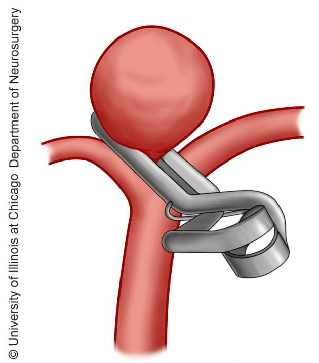
The first and most important step in seeking treatment for a brain aneurysm is to get an expert consultation from an advanced treatment center with expertise in all types of aneurysm treatments, including endovascular therapies and minimally-invasive microsurgical clipping.
Aneurysms that have bled are very serious and considered a medical emergency that requires immediate treatment. In many cases, aneurysm rupture can lead to death or severe disability.
For those fortunate to survive the initial bleeding event, management includes emergent hospitalization to the intensive care unit (ICU) to maintain breathing and vital functions (such as blood pressure) and treatment to reduce brain swelling and to secure the aneurysm to prevent further bleeding.
For patients with an unruptured aneurysm, the risk of a future rupture occurring can be estimated using aneurysm calculators developed from studying a large numbers of patients. If the risk of rupture is greater than the risk of intervention, treatment is recommended in order to prevent rupture and the possible outcomes of death or severe disability.
While endovascular aneurysm treatment is less invasive than microsurgery, whether this is the optimal method to treat an aneurysm depends on the location of the aneurysm, its size and shape, and the patient’s general health.
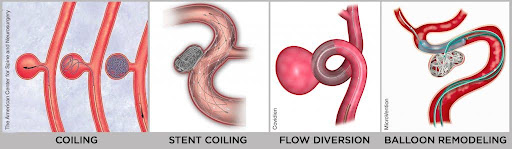
During these procedures, a small tube is inserted into the affected artery and positioned near the aneurysm.
- In aneurysm coiling, specialized metal coils are then moved through the tube into the aneurysm, filling the aneurysm to prevent blood flow into the aneurysm to protect against the risk of rupture. Sometimes the use of a special metal cage called a “stent” can be used to assist the coiling.
- Other times aneurysms can be treated with a specially-designed stent that diverts flow away from the aneurysm (called a flow-diverting stent).
Patient Outcomes
Outcomes for patients with brain aneurysms vary widely depending on factors such as aneurysm size, location, whether it has ruptured, and the timeliness of treatment. Unruptured aneurysms that are diagnosed early and treated effectively generally have excellent prognoses with low complication rates.
For ruptured aneurysms, rapid emergency care is critical to minimize brain damage. Advances in surgical and endovascular techniques have significantly improved survival rates and functional recovery. Rehabilitation services, including physical, occupational, and speech therapy, are often essential parts of recovery for patients who experience neurological deficits.
Meet our Expert Specialists & Surgeons


Experience Compassionate, Expert Care


Written and reviewed by:
The Pacific Neuroscience medical and editorial team
We are a highly specialized team of medical professionals with extensive neurological and cranial disorder knowledge, expertise and writing experience.
Last Updated: