Knowing Your Chordoma Care is in the Right Hands
Superior Treatment for Patients with Clival Chordomas
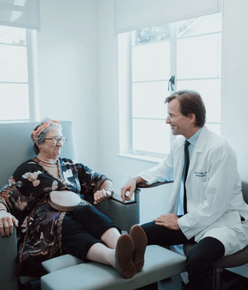
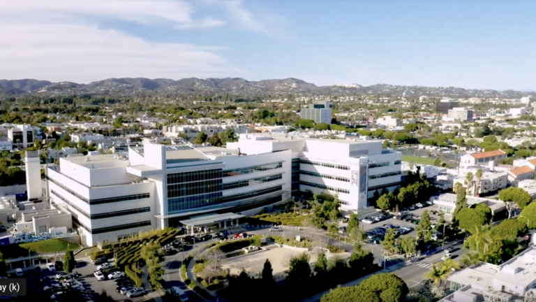
As one of the most comprehensive pituitary disorder programs in the United States, the Pacific Pituitary Disorders Center at Pacific Neuroscience Institute (PNI) offers world-class expert care. Among the top ranked neurology and neurosurgery programs in the nation, our center’s compassionate multidisciplinary specialists provide advanced, personalized treatment while focusing on our patients’ quality of life.
Affiliated with award-winning Providence hospitals Saint John’s Health Center and Little Company of Mary, PNI neurosurgeons lead the way in advancing safer, more effective keyhole and minimally invasive endoscopic pituitary tumor removal approaches.
If you, a family member, or friend have a new diagnosis, require a second opinion, or have a pituitary tumor or related hormonal disorders recurrence, our expert physicians can help you understand your condition and determine an optimal treatment plan.
Think Pituitay. Think PNI.
Contact Us
For information about pituitary disorder treatment please complete the form below. We will respond to you within 12-24 hours. To speak with someone right away contact us at 213-214-2526.
Symptoms
The most common presenting symptom of a clival chordoma is double vision. Less common symptoms may include visual loss, hearing loss, difficulty swallowing, facial numbness, incoordination and motor weakness and nasal congestion.
Diagnosis
These skull base tumors are best diagnosed by MRI and CT scans which will clearly show the extent of tumor and bony destruction. Focused MRIs of the pituitary region, sinuses, temporal bones or internal auditory canals may be indicated to obtain better anatomical detail of a chordoma. Other tests may also be needed prior to surgery such as angiography (typically now performed as a CT angiogram or an MR angiogram), visual field tests, an audiogram or hormonal tests.
Treatment
Initial treatment for a clival chordoma is with surgical removal or debulking. Given their midline location, most clival chordomas and chondrosarcomas are best removed via an endoscopic endonasal approach. However, some extensive and/or laterally placed chordomas may require different skull base surgical approaches. Because chordomas typically invade the bone and dura of the skull base as well as cavernous sinus area, complete surgical resection is often not possible and continued growth of residual tumor is common. Extensive surgery can certainly improve long term survival but over aggressive tumor removal can be associated with significant neurological complications.
Most chordomas are treated after maximal surgical debulking with Stereotactic Radiotherapy (SRT), stereotactic radiosurgery (SRS) or proton beam radiation.
Additionally, while traditional chemotherapy has been relatively ineffective for chordomas, some new targeted agents are now available and showing promise in treating chordomas. At Pacific Brain Tumor Center, by using comprehensive tumor histological subtyping and genomic sequencing, we are able to provide a personalized therapeutic approach for each patient. Our Director of Neuro-Oncology, Dr. Santosh Kesari can facilitate clinical trial participation with novel medical therapies.
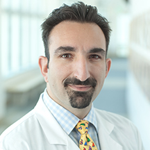
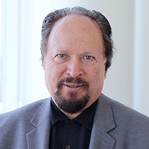
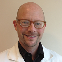
Our Physicians
Click on our award-winning physicians below to learn more about them:


Patient Experience
 Pituitary Disorders Center
 Dennis’ Story – Pituitary Adenoma
Experience an actual account of Pituitary Adenoma.
Dennis’ Story – Pituitary Adenoma
Experience an actual account of Pituitary Adenoma.
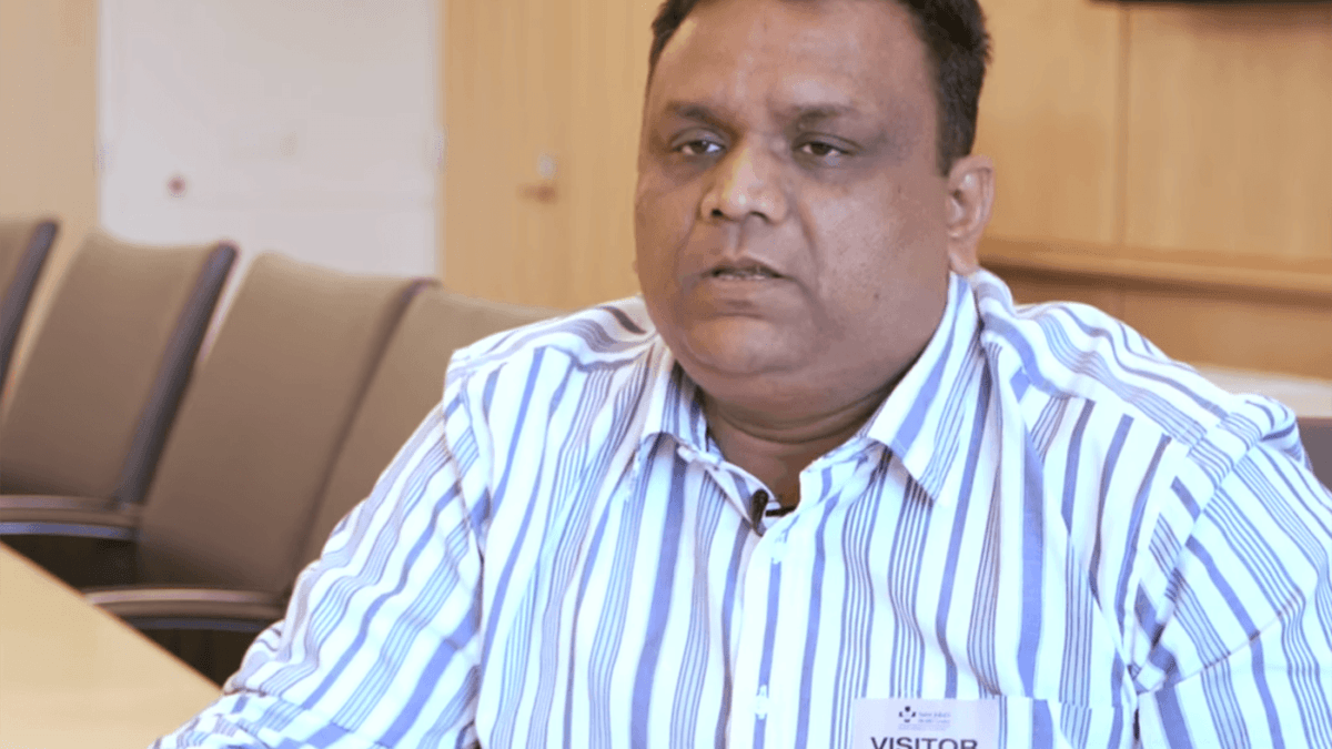 Ajay’s Story – Pituitary Adenoma
Experience an actual account of Pituitary Adenoma.
Ajay’s Story – Pituitary Adenoma
Experience an actual account of Pituitary Adenoma.
Pituitary Disorders Center

Dennis’ Story | Pituitary Adenoma
