Knowing Your Meningioma Care is in the Right Hands
Superior Treatment for Patients with Meningiomas
As one of the most comprehensive brain tumor programs in the United States, the Pacific Brain Tumor Center at Pacific Neuroscience Institute (PNI) offers world-class expert care. Ranked in the top 1% of neurology and neurosurgery programs in the nation, our center’s compassionate multidisciplinary specialists provide advanced, personalized treatment while focusing on our patients’ quality of life.
Affiliated with award-winning Providence hospitals Saint John’s Health Center and Little Company of Mary, PNI neurosurgeons lead the way in advancing safer, more effective keyhole and minimally invasive endoscopic brain tumor removal approaches.
If you, a family member, or friend have a new diagnosis, require a second opinion, or have a brain tumor or skull base tumor recurrence, our expert physicians can help you understand your condition and determine an optimal treatment plan.
Think Brain Tumor. Think PNI.

Contact Us
For information about brain tumor treatment please complete the form below. We will respond to you within 12-24 hours. To speak with someone right away contact us at 213-855-2368.
Symptoms
Midline meningiomas of the skull base that arise above the pituitary gland (tuberculum sellae), or in front of the pituitary gland (planum sphenoidale), typically cause progressive visual loss from optic nerve and optic chiasm compression.
They may also cause:
- Headache
- Double vision
- Loss of pituitary function
Meningiomas that extend into the cavernous sinus may cause double vision (diplopia) and those that extend into the region called Meckel’s cave (below the cavernous sinus), may cause facial numbness or tingling.
Some large petroclival meningiomas that arise in front of the brain stem, may cause:
- Double vision
- Loss of vision
- Facial numbness
- Difficulty walking
- Difficulty swallowing
- Urinary incontinence
- Headache
Diagnosis
What do I do if I have these symptoms?
Meningiomas are best diagnosed by an MRI of the brain with gadolinium, or by a CT scan of the brain with contrast.
Midline skull base meningiomas
For midline skull base meningiomas adjacent to the pituitary gland, a focused MRI of the pituitary region, or orbits is often indicated to obtain better anatomical detail of a meningioma.
Other tests may also be needed such as angiography (a CT angiogram or an MR angiogram), visual field tests, and pituitary hormonal tests.
Treatments
Meningioma Surgery
Symptomatic tuberculum sellae or planum sphenoidale meningiomas are typically treated by surgical removal through either a supraorbital eyebrow or an endoscopic endonasal approach.
Our Center Director, Dr. Daniel Kelly has been one of the pioneers in removing tuberculum sellae meningiomas through an endoscopic endonasal approach and comparing this approach to the supraorbital approach.
Advantage of endoscopic endonasal surgery
The advantage of the endonasal approach over a transcranial approach is that brain retraction is not necessary and manipulation of the optic nerves and chiasm is minimized.
However, for larger tumors (over 3 cms) or those that extend far off the midline, the supraorbital eyebrow approach is an excellent minimally invasive alternative.
With either approach, vision typically improves and pituitary hormonal function is usually preserved. For some larger meningiomas in this region, a more conventional pterional (fronto-temporal) craniotomy may be needed.
For invasive parasellar meningiomas that involve the cavernous sinus, Meckel’s cave, sella and/or petroclival region, endoscopic endonasal tumor debulking and bony decompression is a reasonable treatment option that we often use and often follow with focused stereotactic radiotherapy.
Radiosurgery (SRS) or Stereotactic Radiotherapy (SRT)
Meningiomas of the cavernous sinus, Meckel’s cave, sella tursica and petroclival region cannot be completely removed in over 50% of cases. For meningiomas of the tuberculum sellae or planum sphenoidalea, complete tumor removal rates are significantly higher.
With incomplete removal or if a tumor re-grows stereotactic radiotherapy (SRT) or Stereotactic Radiosurgery (SRS), are often needed to halt further tumor growth.
The tumor control rate with SRS or SRT is quite high with over 90% of patients having no further tumor growth. Complications of SRS or SRT such as visual loss or brain injury are rare.
Our Physicians
Click on our award-winning physicians below to learn more about them:


Patient Experience
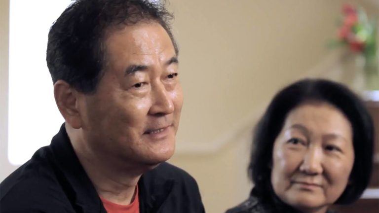
 Akiko’s Story – CNS Lymphoma
Experience Akiko’s recovery from CNS Lymphoma.
Akiko’s Story – CNS Lymphoma
Experience Akiko’s recovery from CNS Lymphoma.
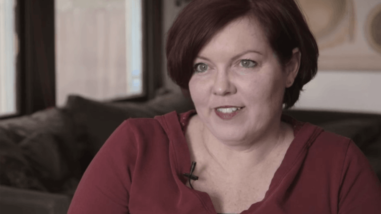
 Justine’s Story – metastatic melanoma
Experience an actual account of metastatic melanoma.
Justine’s Story – metastatic melanoma
Experience an actual account of metastatic melanoma.
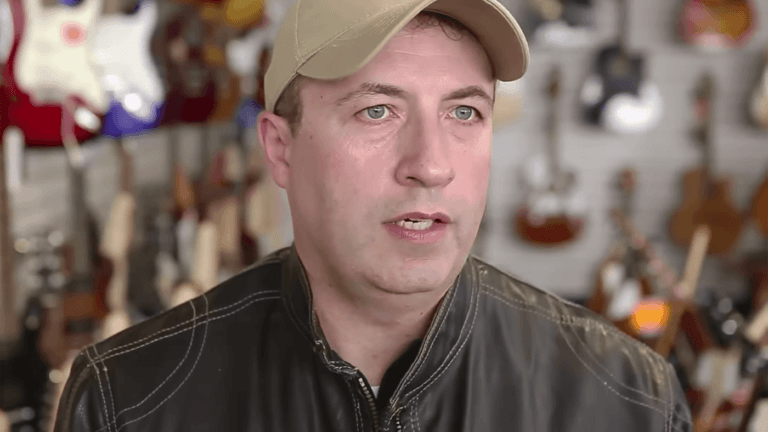
 Chris’ Story- Meningioma Brain Tumor
Experience and actual account of meningioma.
Chris’ Story- Meningioma Brain Tumor
Experience and actual account of meningioma.
 The Pacific Brain Tumor Center
 Meet Dr. Daniel F. Kelly
Akiko’s Story | CNS Lymphoma
Justine’s Story | Metastatic Melanoma
Chris’ Story | Meningioma

The Pacific Brain Tumor Center