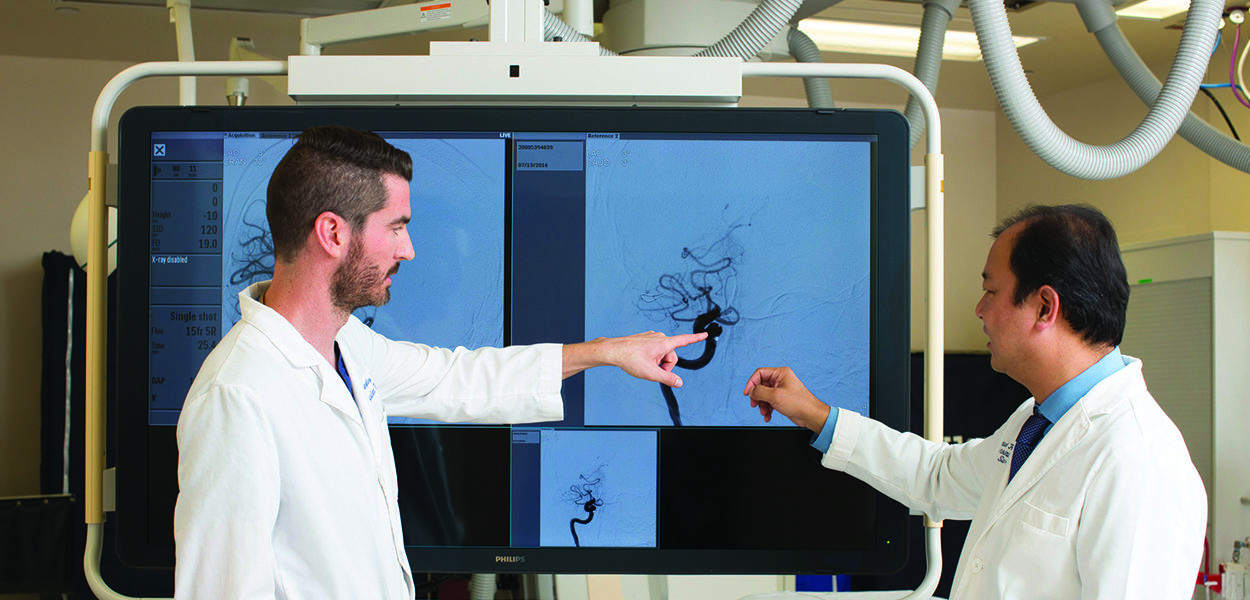
Cavernoma Program
The Cavernoma Program specializes in advanced diagnostics to optimize treatment decisions, including state-of-the-art brain imaging and genetic profiling of vascular malformations to identify targets for therapeutic intervention.
A cavernoma, also called a cavernous malformation (CM), is a vascular abnormality generally found in the brain or spinal cord. Made up of a cluster of abnormal, dilated vessels, blood flows slowly and pools in these “caverns” causing it to appear red to purple in color and have the appearance of a raspberry. Cavernomas contain blood products at various stages of evolution and are usually less than 3 centimeters in size.
Our goal at the Cavernoma Program is to optimize medical management and minimize the need for surgery wherever possible. When refractory cavernomas do progress to require surgical removal, we offer leading-edge minimally invasive microvascular techniques, championing “keyhole” approaches. Benefits include safe corridors that minimize damage to surrounding brain tissues through smaller incisions that reduce post-operative discomfort, faster healing and recovery, and better cosmetic results. Our expert team of neurosurgeons, neuroscientists, vascular biologists and geneticists are leaders in the field of precision therapy for cavernomas, working together to offer patients multidisciplinary team-based care.
If you, a family member, or a friend has been diagnosed with a cavernoma or other vascular malformation, our team of experts is here to help you understand your condition and determine an optimal, personalized treatment plan for your needs. Our aim is to provide our patients with a connected experience of care built on clinical and research excellence.
While a new diagnosis of brain cavernoma can produce natural anxious responses in many patients, the most important thing to remember is that you are not alone — cavernomas can occur in as many as 1 out of every 200 people – and our team of experts is here to help you understand your condition and determine an optimal, personalized treatment plan for your needs.
We use state-of-the-art brain imaging techniques (called diffusion tensor tractography, gradient echo and susceptibility weighted sequences) that allow for noninvasive and computational treatment planning. Most cavernomas only need to be observed with routine brain imaging, repeated over time to observe for changes in appearance, recent bleeding (“hemorrhage”) or appearance of new lesions. Medications are optimized in a patient-specific manner to treat seizures and headaches caused by cavernomas. The Cavernoma Program is also a leader in advanced research initiatives to develop targeted molecular therapies with the goal of preventing new cavernoma formation and protecting against symptomatic bleeding.
If however, refractory cavernomas progress despite optimal medical therapy to require safe surgical removal, our neurosurgery team is specialized in minimally-invasive microvascular techniques with “keyhole” approaches that minimize damage to surrounding brain tissues through smaller incisions that reduce post-operative discomfort and provide better cosmetic results.
Cavernoma diagnosis and treatment
Learn more about our Cavernoma research
Please contact the clinics of neurosurgeon Dr. Garni Barkhoudarian for a consultation or second opinion at 310-582-7450.
