Knowing Your Pituitary Adenoma Care is in the Right Hands
Superior Treatment for Patients with Pituitary Adenomas
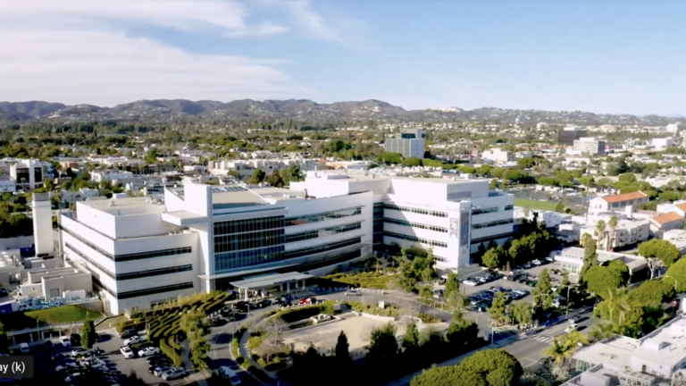
As one of the most comprehensive pituitary disorder programs in the United States, the Pacific Pituitary Disorders Center at Pacific Neuroscience Institute (PNI) offers world-class expert care. Among the top ranked neurology and neurosurgery programs in the nation, our center’s compassionate multidisciplinary specialists provide advanced, personalized treatment while focusing on our patients’ quality of life.
Affiliated with award-winning Providence hospitals Saint John’s Health Center and Little Company of Mary, PNI neurosurgeons lead the way in advancing safer, more effective keyhole and minimally invasive endoscopic pituitary tumor removal approaches.
If you, a family member, or friend have a new diagnosis, require a second opinion, or have a pituitary tumor or related hormonal disorders recurrence, our expert physicians can help you understand your condition and determine an optimal treatment plan.
Think Pituitay. Think PNI.

Contact Us
For information about pituitary disorder treatment please complete the form below. We will respond to you within 12-24 hours. To speak with someone right away contact us at 213-214-2526.
Symptoms
Pituitary adenomas may cause problems because of hormonal hypersecretion, pituitary hormonal failure, vision loss, headaches and/or bleeding into the tumor (apoplexy).
Hormonal hypersecretion
The three most common hormonally active adenomas are prolactinomas, GH-secreting tumors causing acromegaly, and ACTH-secreting tumors causing Cushing’s disease. Thyroid stimulating hormone (TSH) tumors are relatively rare.
Pituitary hormonal deficiency (Hypopitutiarism)
(Hypopituitarism) This problem typically occurs only in larger tumors (macroadenomas) and results from compression and damage to the normal pituitary gland from the enlarging adenoma.
Manifestations may include:
- Hypogonadism – sexual dysfunction, loss of libido, and impotence
- Hypothyroidism – fatigue, weakness, weight gain, coarse dry hair and dry skin, cold intolerance, depression
- Adrenal insufficiency – fatigue, weakness, loss of appetite, dizziness, nausea and vomiting
- Growth failure – in children and adolescents
- Hyperprolactinemia – due to “stalk effect”. This is seen in diseases within or near the pituitary gland and stalk. Interfering with the delivery of dopamine, a neuron-transmitter, from the hypothalamus to the prolactin secreting cells of the pituitary gland), which can result in hypogonadism and its associated problems. Rarely posterior pituitary gland damage occurs with diabetes insipidus, which is caused by the inability of the kidneys to conserve water, leading to frequent urination and thirst.
Neurological problems
Larger pituitary macroadenomas often cause loss of visual acuity or peripheral vision (termed bitemporal hemianopsia) from pressure on the optic nerves and optic chiasm which is directly above the pituitary gland. Larger macroadenomas especially those that have hemorrhaged (apoplexy) may also cause double vision.
Headache
Patients with macroadenomas often have frontal, forehead and temporal area headaches. Pituitary adenoma removal often results in headache resolution.
Bleeding (pituitary apoplexy)
This condition develops over hours to several days from hemorrhage and/or infarction of pituitary adenoma (typically a macroadenoma or a Rathke’s cleft cyst).
Symptoms may include:
- Headache
- Nausea
- Visual loss
- Double vision
- Confusion
Most patients have undiagnosed hormone insufficiency prior to the apoplectic event. Pituitary apoplexy is best confirmed by MRI. A head CT will also show areas of bleeding or a mass in the sella in the majority of cases. Other conditions that can mimic pituitary apoplexy are a ruptured aneurysm, meningitis, a stroke, intracerebral hemorrhage and migraine headache.
The treatment for most patients with pituitary apoplexy is urgent endoscopic endonasal surgery and intravenous administration of the stress hormone cortisol.
Diagnosis
Pituitary adenomas are best diagnosed by imaging studies and hormonal testing.
Imaging
The imaging study of choice is an MRI of the pituitary gland without and with gadolinium (a contrast agent). A brain MRI or CT scan will also reveal most pituitary macroadenomas but may not reveal smaller microadenomas.
In a minority of patients it may be difficult to distinguish an adenoma of the pituitary from other masses which may include:
- Craniopharyngioma
- Rathke’s Cleft cyst
- Meningioma
- Hypophysitis (pituitary inflammation)
- Glioma of the suprasellar region
- Metastatic tumor
- Cordoma
Pituitary Hormonal Testing
Evaluation and interpretation of the pituitary gland function either for hormonal deficits or inappropriate hormonal secretion is performed in our center, under the supervision of our pituitary neuroendocrinologist Dr. Katherine Araque.
Our team recognizes that one size does not fit all. Consequently, pituitary hormonal testing is individualized to the needs of our patients.
Our center can be easily perform these types of blood level measurements:
- ACTH
- Serum cortisol
- TSH
- Free T4
- Total T3 (thyroid function)
- LH (luteinizing hormone)
- FSH (follicle-stimulating hormone)
- Estradiol in women
- Free and total testosterone in men
- GH (growth hormone)
- IGF-1 (insulin-like growth factor)
- Prolactin
- Macroprolactin
Additionally, patients can have access to various diagnostic tools including radiologic images with selected pituitary protocols, noninvasive and invasive dynamic endocrinology testing.
Neuro-Ophthalmological Evaluation
Patients with visual complaints or those whose tumors that contact the optic nerves or optic chiasm should receive a full ophthalmological evaluation. An evaluation with our neuro-ophthalmologist, Dr. Howard Krauss, should include acuity (vision quality) testing of each eye and formal visual field testing to determine if there is loss of peripheral vision.
Treatment
Pituitary Adenoma (Tumor) Surgery
Endoscopic Endonasal Surgery
The first-line treatment for all pituitary adenomas (tumors) except prolactinomas (as discussed below), as well as Rathke’s Cleft Cysts (RCCs) and most craniopharyngiomas is endoscopic endonasal transsphenoidal surgery. Because of improved tumor visualization and tumor removal rates, the endoscopic endonasal approach has become the preferred method for removal of pituitary adenomas, RCCs, as well as the great majority of craniopharyngiomas, clival chordomas and many midline meningiomas.
Surgical success rates (complete tumor removal and normalization of hormonal hypersecretion) are generally quite high (>80-90%) with smaller and non-invasive pituitary adenomas, but are lower for large or invasive macroadenomas (40-70%).
Major surgical complications such as vision loss, bleeding, stroke, cerebrospinal fluid leak and meningitis are low when performed by experienced endonasal transsphenoidal neurosurgeons who often work collaboratively with a Head & Neck surgeon. The success rates of surgery are described further in the sections below for specific tumor types.
Transcranial Surgery
A craniotomy (opening in the skull from above to reach the pituitary region) is required in less than 1% of patients who require surgery for a pituitary adenoma.
Medical & Radiation Therapies
See specific pituitary adenoma subtypes:
- Acromegaly
- Cushing’s disease
- Prolactinoma
- TSH-secreting
- Endocrine-inactive
Hormone Replacement Therapy
Many patients with pituitary adenomas will develop varying degrees of hypopituitarism (pituitary gland deficiency) either as a result of the tumor impact on the gland or sometimes as a result of surgery or other treatments. While many patients can have some degree of gland recovery, many will ultimately require short-term or long-term hormone replacement therapy. Our highly experienced pituitary endocrinologists, Drs. Pejman Cohan and Katherine Araque can provide tailored hormone replacement therapy including for low testosterone, low estrogen, growth hormone deficiency, hypothyroidism, low cortisol (adrenal insufficiency) and diabetes insipidus (anti-diuretic hormone deficiency).
For prolactinomas that are typically treated with medical therapy, we have highly experienced endocrinologists to provide this therapy. For patients with adenomas that cannot be completely treated with surgery and/or medical therapy, our experienced radiation oncologists experts can deliver focused radiosurgery or radiotherapy to tumors, halting their growth.
We also have clinical trials available using novel medical therapies for patients with persistent acromegaly and Cushing’s disease who failed prior treatments.
Our Physicians
Click on our award-winning physicians below to learn more about them:


Patient Experience
 Pituitary Disorders Center
 Dennis’ Story – Pituitary Adenoma
Experience an actual account of Pituitary Adenoma.
Dennis’ Story – Pituitary Adenoma
Experience an actual account of Pituitary Adenoma.
 Ajay’s Story – Pituitary Adenoma
Experience an actual account of Pituitary Adenoma.
Ajay’s Story – Pituitary Adenoma
Experience an actual account of Pituitary Adenoma.
Pituitary Disorders Center

Dennis’ Story | Pituitary Adenoma
