Knowing Your Glioma Care is in the Right Hands
Superior Treatment for Patients with Gliomas
As one of the most comprehensive brain tumor programs in the United States, the Pacific Brain Tumor Center at Pacific Neuroscience Institute (PNI) offers world-class expert care. Ranked in the top 1% of neurology and neurosurgery programs in the nation, our center’s compassionate multidisciplinary specialists provide advanced, personalized treatment while focusing on our patients’ quality of life.
Affiliated with award-winning Providence hospitals Saint John’s Health Center and Little Company of Mary, PNI neurosurgeons lead the way in advancing safer, more effective keyhole and minimally invasive endoscopic brain tumor removal approaches.
If you, a family member, or friend have a new diagnosis, require a second opinion, or have a brain tumor or skull base tumor recurrence, our expert physicians can help you understand your condition and determine an optimal treatment plan.
Think Brain Tumor. Think PNI.

Contact Us
For information about brain tumor treatment please complete the form below. We will respond to you within 12-24 hours. To speak with someone right away contact us at 213-855-2368.
Symptoms
Glioma brain tumor symptoms are variable and depend on tumor location and size.
The first symptom of a brain tumor of any type can be a headache. The reason that patients get headaches with brain tumors is that these masses cause increased pressure in the brain. The headache associated with a brain tumor is frequently worse in the morning and may be associated with nausea or vomiting.
Other common signs and symptoms of a gliomas include:
- Headache
- Seizures
- Confusion
- Weakness
- Numbness of a side or part of the body
- Imbalance
- In-coordination
- Memory impairment
- Changes in mood
- blurred, double or loss of vision
Diagnosis
What do I do if I have these symptoms?
If a patient has any of the symptoms mentioned above with no other obvious explanation a diagnostic work-up should be done.
Glioma, like other brain tumors, is currently best seen on magnetic resonance imaging (MRI) studies of the brain. Computerized tomography (CT) is also a good technique for seeing structures in the head and brain but does not show quite as much detail, as does MRI.
The problem with MRI is that while it is a good technique for detecting a brain mass, it does not identify the type of mass. A glioma can look like other kinds of brain tumor, or even like an infection. A resection is needed to confirm the diagnosis of glioma and to grade the tumor, in order to guide the next steps of appropriate treatment and provide a prognosis for survival time.
The microscopic structure of a glioma is extremely important in making decisions about treatment and in predicting survival. Based on features of cells within tumors, neuropathologists can often predict how aggressively a tumor will behave, and assign a WHO grade, which ultimately contributes to how long a patient will survive and what quality of life they may have.
Treatments
The treatment of a glioma depends tremendously on the tumor WHO grade and subtype, as well as size and location of the tumor. Through many decades of research, we have learned that for most gliomas, the amount of tumor removed may correlate with survival time. At the same time, for higher grade (WHO Grade III and IV) gliomas including glioblastoma multiforme, although all visible portions of the tumor may be removed with surgery, the tumor has often already infiltrated the surrounding brain far beyond the margins of the visible tumor, making a surgical cure impossible.
Because gliomas are infiltrative into the brain and blend into the adjacent normal brain, they typically cannot be removed completely.
Glioma Brain Tumor Surgery
Treatment for gliomas can be surgical or non-surgical. One of the key principles we focus on is maximal tumor removal with preservation of quality of life and neurological function. Typically through a keyhole craniotomy approach followed by radiotherapy and chemotherapy. In many instances, all 3 of these treatments are needed. To maintain this standard, we offer the most advanced, state-of-the-art, surgical techniques for glioma removal, including:
- Neurophysiological monitoring
- Standard Motor Mapping
- Advanced neuro-imaging (functional MRI, MR perfusion, tractography, spectroscopy)
- Awake speech and motor mapping
- Neuro-navigation technology
Radiation Therapy for Gliomas
Radiation therapy is an effective treatment option for most gliomas. Radiation can be administered to the whole brain, or it can be relatively focused to a selected region of the brain via a technology called radiosurgery.
PNI offers the benefits of several types of radiation therapy, both of which allow very precise focusing of radiation beams into the area of glioma with minimized damage to surrounding brain.
Chemotherapy for Gliomas
Chemotherapy is another option for treatment of gliomas. The current standard of therapy for newly-diagnosed high grade gliomas (WHO Grade III or IV) typically include 6 weeks of chemotherapy using a drug called temozolamide, in conjunction with radiation therapy followed by additional chemotherapy cycles.
Many patients will have the option of enrolling in a major clinical trial, typically investigating an experimental intervention for treating these tumors. Clinical trials for glioblastoma are essential to fighting this disease. Personalized therapy options based on molecular and genetic profiling of the tumor also allow PNI to determine treatments that are individualized for each patient.
Our Physicians
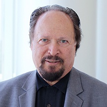
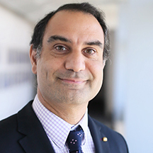
Click on our award-winning physicians below to learn more about them:
Patient Experience
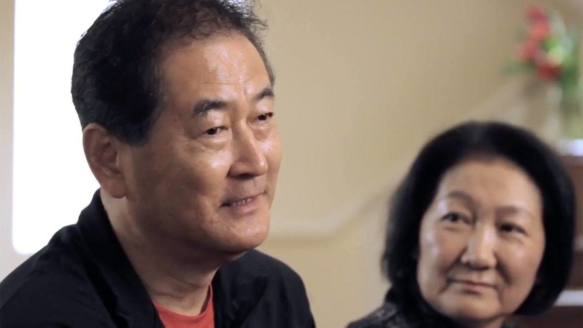 Akiko’s Story – CNS Lymphoma
Experience Akiko’s recovery from CNS Lymphoma.
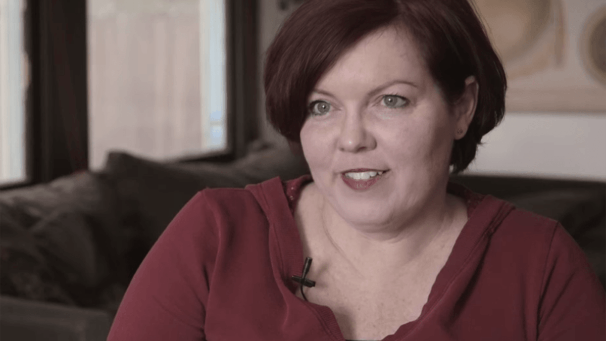 Justine’s Story – metastatic melanoma
Experience an actual account of metastatic melanoma.
 Chris’ Story- Meningioma Brain Tumor
Experience and actual account of meningioma.
 The Pacific Brain Tumor Center
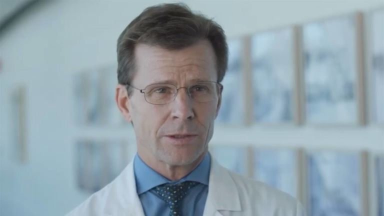
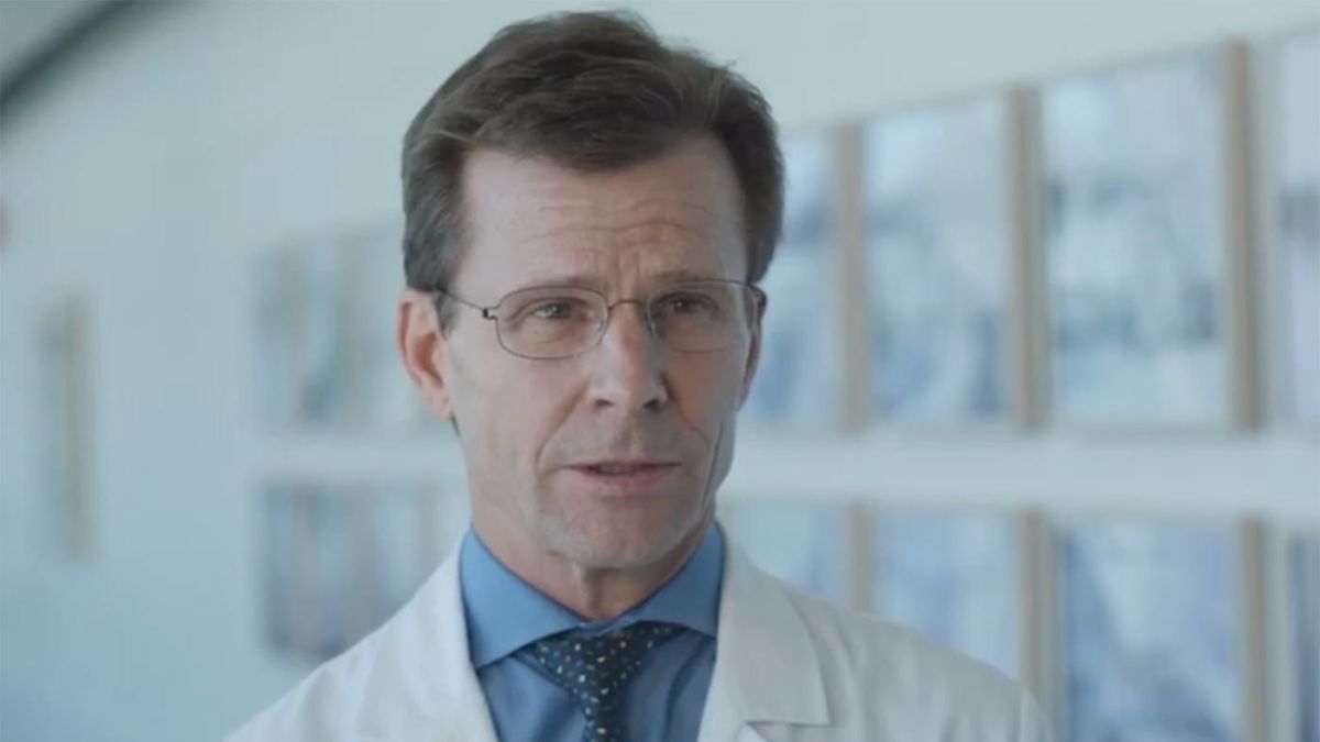 Meet Dr. Daniel F. Kelly
Akiko’s Story | CNS Lymphoma
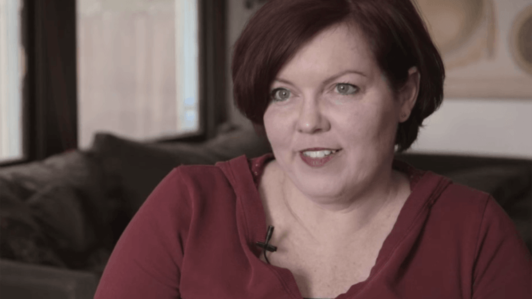
Justine’s Story | Metastatic Melanoma
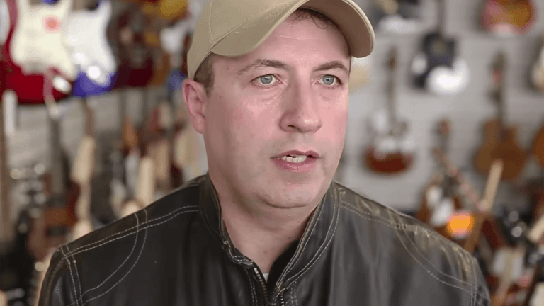
Chris’ Story | Meningioma

The Pacific Brain Tumor Center
