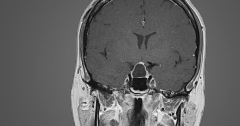
Sellar Arachnoid Cyst
What are Sellar Arachnoid Cysts?
Sellar arachnoid cysts are benign fluid-filled masses that can cause loss of pituitary gland function, visual loss & headache. Arachnoid cysts occurring in the sella and suprasellar region are relatively uncommon. Like arachnoid cysts in other parts of the intracranial space, sellar arachnoid cysts are thought to develop from an arachnoid* pocket or diverticulum that progressively fills with cerebrospinal fluid (CSF).
As these benign cysts enlarge, the can put pressure on the adjacent pituitary gland, optic chiasm and nerves and surrounding skull base membranes, leading to loss of pituitary gland function, visual loll and/or headaches.
Diagnosis of Sellar Arachnoid Cysts
Accurate diagnosis of sellar arachnoid cysts involves a comprehensive evaluation by a multidisciplinary team of neurosurgeons, neurologists, and neuroradiologists. The diagnostic process typically includes the following steps:
- Medical History and Physical Examination: Your healthcare provider will begin by conducting a detailed medical history interview, gathering information about your symptoms, medical background, and any relevant family history. A comprehensive physical examination will also be performed to assess any neurological signs associated with the cyst.
- Neuroimaging Studies: To visualize and evaluate the sellar region, various neuroimaging techniques are employed. Magnetic Resonance Imaging (MRI) is the primary imaging modality used to identify and characterize sellar arachnoid cysts. It provides detailed cross-sectional images of the brain, allowing our experts to assess the location, size, and impact of the cyst on surrounding structures.
- Hormonal Evaluation: Since sellar arachnoid cysts can potentially affect pituitary gland function, hormone level testing may be conducted to assess any hormonal imbalances or deficiencies. Endocrine specialists collaborate closely with our team to analyze these results and provide valuable insights into the overall hormonal status.
- Collaborative Multidisciplinary Evaluation: At Pacific Neuroscience Institute, we follow a collaborative approach to diagnosing sellar arachnoid cysts. Our team of experts, including neurosurgeons, neurologists, neuroradiologists, and endocrinologists, carefully review all clinical and diagnostic findings to arrive at an accurate diagnosis. This multidisciplinary evaluation ensures comprehensive assessment and optimal treatment planning tailored to your specific needs.
Treatment of Sellar Arachnoid Cysts
Treatment of symptomatic sellar and suprasellar arachnoid cysts is ideally performed through an endoscopic endonasal approach using a simplified cyst obliteration technique with an abdominal fat graft. Sellar arachnoid cysts can be treated with simple endoscopic cyst obliteration – a technique described by our group
This technique was originally described by our group in the Journal of Neurosurgery in 2012 and was shown to have a high success rate and low complication rate.
At Pacific Pituitary Disorders Program located in Santa Monica, CA, we have one of the world’s largest experiences in transsphenoidal endoscopic endonasal surgery. By incorporating cutting edge technology and instrumentation with proven surgical experience of over 1600 endonasal surgeries, we make arachnoid cyst surgery safer, less invasive and more effective.
*Arachnoid is a thin delicate membrane that surrounds the brain and spinal cord.


